Coping with Cancer
Log in / Sign up
Forum

Together
We Find
Strength

Explore what we offer >>

Slide 1
No matter
what the
Statistics say
there is
always a
Welcome to copingwithcancer.in
Way Ahead
Slide 2
No matter
what the
Statistics say
there is
always a
Info Hub for
Cancer patients & survivors
Way Ahead
Slide 3
No matter
what the
Statistics say
there is
always a
All the Help
You need for the Fight ahead
Way Ahead

Category: New Frontiers in Cancer Research

  • Astrocytes’ hidden potential: Cross-species study reveals new insights into spinal cord injury repair

    Astrocytes’ hidden potential: Cross-species study reveals new insights into spinal cord injury repair

    Spinal cord injury (SCI) remains one of the most devastating medical conditions, severely impacting quality of life and often leading to permanent disability. The central nervous system (CNS) has a limited capacity for regeneration, which poses a significant challenge in treating SCI, since recovery becomes increasingly difficult once the spinal cord is damaged.

    During development, neural stem cells in the spinal cord differentiate into various neural cells that form complex circuits. As the spinal cord matures, these progenitor cells lose their regenerative potential, making adult spinal cord tissue less capable of recovery after injury. Identifying endogenous stem cells with diverse lineage potentials in the adult spinal cord is crucial for advancing repair strategies for SCI.

    In a recent study published in the Proceedings of the National Academy of Sciences, researchers led by Profs. Dai Jianwu and Zhao Yannan from the Institute of Genetics and Developmental Biology of the Chinese Academy of Sciences explored the regenerative mechanisms behind SCI repair.

    By constructing single-cell transcriptomic databases for both human spinal cord development and rhesus monkey SCI models, the researchers established a foundation for comprehensively analyzing spinal cord cell behavior. Their cross-species examination of primate and rodent spinal cords revealed new insights into the behaviors of ependymal and astrocyte cells following SCI, highlighting their dynamic roles in spinal cord repair.

    The research team discovered that, during spinal cord development, ependymal cells mature and gradually lose their neural progenitor cell properties, retaining only limited proliferative capacity. After injury, ependymal cells displayed minimal activation and demonstrated no significant proliferation or cross-lineage differentiation.

    Importantly, the study revealed that the reactivity of ependymal cells post-SCI is significantly lower in primates than in rodents, indicating more restricted regenerative potential in primates.

    In contrast, astrocytes in the injured spinal cord exhibited significant activation. Through single-cell and lineage tracing analyses, the researchers revealed that some astrocytes could transdifferentiate into oligodendrocytes under injury-induced conditions, thereby contributing to the remyelination process.

    Further investigation identified an intermediate population among astrocytes where key transcription factors such as SOX10 promoted their conversion into oligodendrocyte lineage cells.

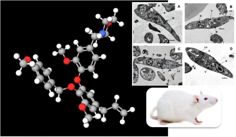
    To enhance the regenerative process, the team introduced functional material transplantation into the injury microenvironment. This intervention significantly boosted the efficiency of astrocyte transdifferentiation into oligodendrocytes, suggesting that material transplantation not only mitigates the inhibitory effects of the injury site but also creates favorable conditions for promoting remyelination.

    This study provides compelling evidence of the limited regenerative capacity of ependymal cells in adult primate spinal cord injury and underscores the transdifferentiation potential of astrocytes. Furthermore, the research highlights how microenvironmental modulation can enhance the efficiency of astrocyte-mediated repair, offering a promising approach for future SCI therapies.

  • Potent compound derived from Brazilian plant offers hope for neglected disease

    Potent compound derived from Brazilian plant offers hope for neglected disease

    A compound derived from Nectandra leucantha, a tree native to southern Brazil (local names: canela-seca or canela-branca), has the potential to be used to treat visceral leishmaniasis, a neglected tropical disease associated with poverty, malnutrition, poor housing and lack of basic sanitation.

    The disease is almost always fatal if left untreated. Most cases occur in Brazil, East Africa and India, according to the World Health Organization (WHO). An estimated 50,000–90,000 new cases and 20,000–50,000 deaths occur worldwide annually, with only 25%–45% of cases being reported to WHO.

    The disease is caused by a protozoan parasite transmitted by a sandfly bite, and is characterized by long bouts of fever, loss of weight and muscle strength, enlargement of the spleen and liver, and anemia.

    An article by researchers affiliated with institutions in Brazil, the United Kingdom and Portugal, published in the journal Antimicrobial Agents and Chemotherapy, reports the findings of a study showing that the substance killed Leishmania infantum, the parasite that causes the disease, selectively (i.e. without affecting host cells).

    The first step in the study was synthesis of a compound similar to dehydrodieugenol B, a neolignan found naturally in N. leucantha and isolated originally by João Lago, full professor at the Federal University of the ABC (UFABC) in São Paulo state, Brazil. The synthesis was performed by Edward Anderson, a professor of organic chemistry at the University of Oxford in the UK.

    “We used this substance as a prototype, a model based on which we could design novel versions of the molecule [with minor structural variations] and test them one by one on the parasite in vitro with the aim of optimizing its action,” said André Gustavo Tempone, principal investigator for the study and a researcher at Butantan Institute’s Physiopathology Laboratory in Brazil.

    In this manner, the researchers obtained a molecule four times more powerful than the prototype. However, in vivo tests involving animals were disappointing because the optimized compound circulated in the rodents’ organism for less than ten minutes, and the study was unable to make progress.

    “The fact that the substance circulated for such a short time in the rats’ bodies suggested that the ensuing stages of the research would fail. It became clear that the substance wouldn’t produce the expected results,” Tempone said.

    The team then focused on additional optimization of the molecule with the aim of enhancing its bioavailability so that it would remain for longer in the animal’s organism. After several chemical optimization processes conducted in vitro in partnership with Maiara Amaral, a student of Tempone who was on an internship at Oxford University and used the project as her Ph.D. thesis, they arrived at a more potent molecule whose mean plasma half-life reached 21 hours.

    Pharmacokinetic studies measuring the time required for the substance to be absorbed, distributed, metabolized and excreted showed that it circulated in the rat’s organism for a period 100 times longer than that observed initially.

    Based on the in vitro analysis, the researchers concluded both that the novel substance was more potent in combating L. infantum, and that it did no damage to host cells. They also investigated its action mechanism, showing that it caused an irreversible collapse of the parasite’s energy mechanism (ATP) due to an increase in calcium, while reducing host cell inflammation, a key factor in the treatment of visceral leishmaniasis. With these good results behind them, the scientists plan to advance farther in animal trials.

    “We need to analyze the action of the compound in rodents with leishmaniasis in order to assess its efficacy and the doses required for treatment,” Tempone said.

    Their long-term goal is to use the compound to produce medications against visceral leishmaniasis, but a great deal of work still has to be done to achieve this objective. As Tempone recalled, novel drugs take around 15 years to come to market, the development process involving rigorous tests and trials to ensure that the active ingredient is totally safe before clinical trials involving humans can be approved.

    This research is extremely important, he added, as the large pharmaceutical companies are not interested in developing drugs for neglected diseases such as visceral leishmaniasis.

    “Brazil has one of the most outstanding biodiversities in the world, and a huge abundance of available chemical structures that can be copied and used in medications. If we don’t invest in combating this disease, the rich countries where it isn’t endemic certainly won’t,” he said.